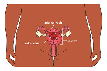
Adenomyosis is a condition where cells similar to those that line the uterus are also in the muscle wall of your uterus. Studies suggest that about one in five women have adenomyosis. Learn more about the symptoms, causes, diagnosis and treatments.
What is adenomyosis?
Symptoms
Causes
Diagnosis
Fertility and pregnancy
Treatment and management
When to see your doctor
Related resources
Adenomyosis is a condition where cells similar to those that line the uterus are also in the muscle wall of your uterus (usually in the back wall).
If adenomyosis is mainly in one area, it can lead to a noncancerous growth called an ‘adenomyoma’.

Women with adenomyosis often have endometriosis too, but the conditions are different. With endometriosis, the cells are found on other parts of your body such as your fallopian tubes, ovaries or tissue lining your pelvis.
With adenomyosis, the cells in the muscle wall behave the same way as cells lining the uterus. When you have your period, the cells in the muscle wall also bleed. But because they are trapped in the muscle layer, they form little pockets of blood in the muscle wall.
About two-thirds of women with adenomyosis experience:
We don’t know the exact cause of adenomyosis, but it may be associated with:
Adenomyosis only occurs in women who have periods, particularly women aged 30 to 50.
You doctor will ask questions about your symptoms and may ask to do a vaginal examination. If you have adenomyosis, your uterus might feel tender or enlarged.
Adenomyosis can be hard to diagnose because there are no agreed tests to confirm the condition.
Your doctor might refer you to a gynaecologist. Depending on your situation, they may do a transvaginal ultrasound (an ultrasound probe is gently placed into your vagina).
You may also need an MRI scan to exclude other conditions, such as fibroids, and confirm the diagnosis.
Adenomyosis cannot be diagnosed from blood tests or tissue samples (biopsies). Diagnosis is often only confirmed with pathology tests after the uterus has been removed (hysterectomy).
Adenomyosis can cause fertility problems because the condition makes it hard for an embryo to implant into the uterine lining.
There may also be pregnancy complications, and anaemia from heavy bleeding.
Treatment for adenomyosis will depend on your symptoms, stage of life and whether you plan to have children. You can also try things like gentle physical activity, meditation, yoga and acupuncture in addition to standard treatments to help manage your symptoms.
Research shows the Mirena® IUD has the best outcomes for managing symptoms of adenomyosis. The IUD is inserted into your uterus. It releases progesterone, which reduces bleeding and pain and thins the endometrial cells. You can have the IUD removed if you are planning a pregnancy.
The combined oral contraceptive pill may reduce bleeding and pain, but research suggests it is not as effective as the Mirena® IUD. You can stop taking either treatment if you are planning a pregnancy.
Your doctor or specialist may not recommend surgery if you are planning a pregnancy. Surgery can result in scar tissue in the uterus, which might affect your fertility.
If you are not planning any future pregnancies, you can have an operation to remove the uterus lining (endometrial ablation) to reduce heavy bleeding. This operation may not reduce pain. You can also combine an endometrial ablation with an IUD.
If your symptoms stop you from doing normal activities – and you don’t want to have a child or more children – you can have an operation to remove your uterus (hysterectomy).
Uterine artery embolisation is a non-surgical procedure that blocks blood supply to part of the uterus. This procedure reduces pain and bleeding. But it is not recommended if you are planning a future pregnancy.
Talk to your doctor if your symptoms (physical and emotional) stop you from doing things you normally do.
This content has been reviewed by a group of medical subject matter experts, in accordance with Jean Hailes policy.
© Jean Hailes Foundation. All rights reserved.